• Skip to main content
  • Skip to primary sidebar
  • Skip to footer

Materials Characterization Facility

  • About
    • Staff
    • Photo Gallery
  • Instruments
    • Equipment Status
    • Fees
    • Forms
    • MSDS
    • Policies
  • Training
  • Reservations
You are here: Home / Instruments / Dimension Icon AFM

Dimension Icon AFM

Dimension Icon AFM

The MCF has a Bruker Dimension Icon AFM for scanning probe microscopy. With an atomic force microscope (AFM), a sharp probe is scanned in a raster pattern across the sample surface to obtain information such as topography in  three dimensions. Other information such as magnetic properties, chemical properties, or forces between the tip and the sample can also be measured using AFM.  Since the AFM’s resolution is not limited by the diffraction of light, it can be used to achieve high resolution (Z <0.1 nm; XY <1 nm).

Dimension Icon AFM capabilities:

  • Peak Force Tapping using ScanAsyst: Topographs and inphase images.
  • Contact mode atomic force microscopy (CM-AFM): Topographs and lateral force frames.
  • Force Imaging: Elastic properties of materials from force curves plots.
  • Intermittent mode (tapping) atomic force microscopy (TM-AFM): Topographs and phase images.
  • Imaging in a liquid environment: Imaging in liquid can be accomplished using ScanAsyst, tapping or contact mode AFM.
  • Peak Force TUNA: Topographs, Current images, current-voltage (I-V) plots.
  • Peak Force Quantitative NanoMechanics (PF-QNM): Modulus, adhesion, deformation and dissipation measurements.
  • Magnetic Force Microscopy (MFM): long range magnetic forces on the sample surface.

Icon AFM Instructions (updated 9/4/2012)

For more information, go to www.bruker.com

For training on this instrument, please contact Dr. Wilson Serem.

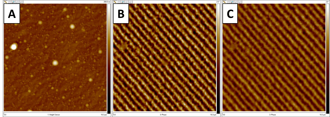
Magnetic domains of a magnetic recording tape mapped using magnetic force microscopy (MFM). A) Topography view; B) MFM phase image obtained at scan lift height set at 40nm; C) MFM phase frame obtained at scan lift height set at 80nm. Image taken by Wilson Serem.

Primary Sidebar

Instruments

PI 85 and PI 95 PicoIndenter

AFM-IR

Omicron XPS/UPS

Nanoindenter

LYRA FIB-SEM

Ellipsometer

Spectrofluorometer

UV-Vis-NIR Spectrophotometer

Electopolishers

Confocal Microscope

FE-SEM

Cameca Ion Microprobe

Dimension Icon AFM

LYRA FIB-SEM

Fume Hoods

FERA FIB-SEM

Electron Microprobe

FTIR Spectrometer

Differential Scanning Calorimeter (DSC)

Thermal Mechanical Analysis (TMA)

Dynamic Mechanical Analysis (DMA)

Hot Disc Thermal Conductivity Analyzer

Dielectric Spectrometer

Tensile Stages (500 N and 10 kN)

Prep Lab

Titan Themis 300 S/TEM

Optical Microscope

PIPS II

SEM and TEM Plasma Cleaners

Particle Size Analyzer

Surface Area Analyzer

Footer

Materials Characterization Facility (MCF)

Frederick E. Giesecke Engineering Research Building
1617 Research Parkway
College Station, TX 77843

Texas A&M Experiment Station
  • Accessibility
  • State Links and Policies
  • Privacy Notice
  • Website Feedback
  • Texas A&M University

Copyright © 2023 · Texas A&M Engineering Experiment Station · All Rights Reserved