• Skip to main content
  • Skip to primary sidebar
  • Skip to footer

Materials Characterization Facility

  • About
    • Staff
    • Photo Gallery
  • Instruments
    • Equipment Status
    • Fees
    • Forms
    • MSDS
    • Policies
  • Training
  • Reservations
You are here: Home / Instruments / FE-SEM

FE-SEM

JEOL JSM-7500F

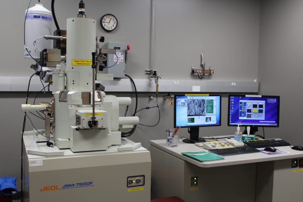
The JEOL JSM-7500F is an ultra high resolution field emission scanning electron microscope (FE-SEM) equipped with a high brightness conical FE gun and a low aberration conical objective lens).

The improved overall stability of the JSM-7500F enables you to readily observe your specimen at magnifications up to 1,000,000x with the guaranteed resolution of 1 nm.

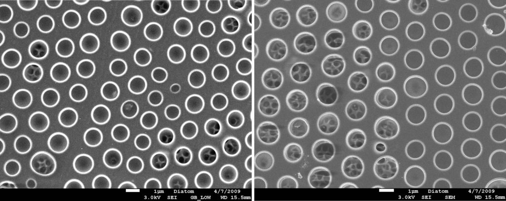
The energy filter (r-filter) makes it possible to observe the fine surface morphology of nanostructures.

The unique Gentle Beam (GB) mode decelerates incident electrons just before they hit the specimen to reduce the incident-electron penetration and the charging in the specimen.  The GB mode provides high-resolution images whose quality is as high as those of higher accelerating voltages, even at low accelerating voltages from 100 V to 3 kV without damaging the specimen surface.

Source: Cold cathode UHV field emission conical anode gun

Resolution: 1.0 nm guaranteed at 15kV; 2.2 nm guaranteed at 1.0kV

Acc. voltage: 0.5 to 30kV (SEM or LM mode); 10 V steps from 0.5 to 2.9 kV; 100 V steps from 2.9 to 30 kV

Mag. range: 25x to 19,000x in LM mode; 100x to 650,000x in SEM mode

Probe current: 1 x 10-13 order to > 2 x 10⁻⁹ Amps (continuous)

Accessories associated with the JSM-7500 include: conventional in-chamber Everhart-Thornley and through-the-lens secondary detectors, low angle back-scattered electron detector (LABE), IR-CCD chamber camera, Oxford EDS system equipped with X-ray mapping and digital imaging.

FE-SEM Instructions

FE-SEM Training Policy

To be trained on this instrument, please contact Dr. Yordanos Bisrat.

Comparison of an uncoated diatom using gentle beam mode (left) versus normal mode (right) in the JEOL JSM-7500F field emission SEM. Note the charging visible on the left side of the image taken in normal mode (blurriness).

Primary Sidebar

Instruments

PI 85 and PI 95 PicoIndenter

AFM-IR

Omicron XPS/UPS

Nanoindenter

LYRA FIB-SEM

Ellipsometer

Spectrofluorometer

UV-Vis-NIR Spectrophotometer

Electopolishers

Confocal Microscope

FE-SEM

Cameca Ion Microprobe

Dimension Icon AFM

LYRA FIB-SEM

Fume Hoods

FERA FIB-SEM

Electron Microprobe

FTIR Spectrometer

Differential Scanning Calorimeter (DSC)

Thermal Mechanical Analysis (TMA)

Hot Disc Thermal Conductivity Analyzer

Dielectric Spectrometer

Tensile Stages (500 N and 10 kN)

Prep Lab

Titan Themis 300 S/TEM

Optical Microscope

PIPS II

SEM and TEM Plasma Cleaners

Particle Size Analyzer

Surface Area Analyzer

Footer

Materials Characterization Facility (MCF)

Frederick E. Giesecke Engineering Research Building
1617 Research Parkway
College Station, TX 77843

Texas A&M Experiment Station
  • Accessibility
  • State Links and Policies
  • Privacy Notice
  • Website Feedback
  • Texas A&M University

Copyright © 2025 · Texas A&M Engineering Experiment Station · All Rights Reserved