• Skip to main content
  • Skip to primary sidebar
  • Skip to footer

Materials Characterization Facility

  • About
    • Staff
    • Photo Gallery
  • Instruments
    • Equipment Status
    • Fees
    • Forms
    • MSDS
    • Policies
  • Training
  • Reservations
You are here: Home / Instruments / Confocal Microscope

Confocal Microscope

Confocal Microscope

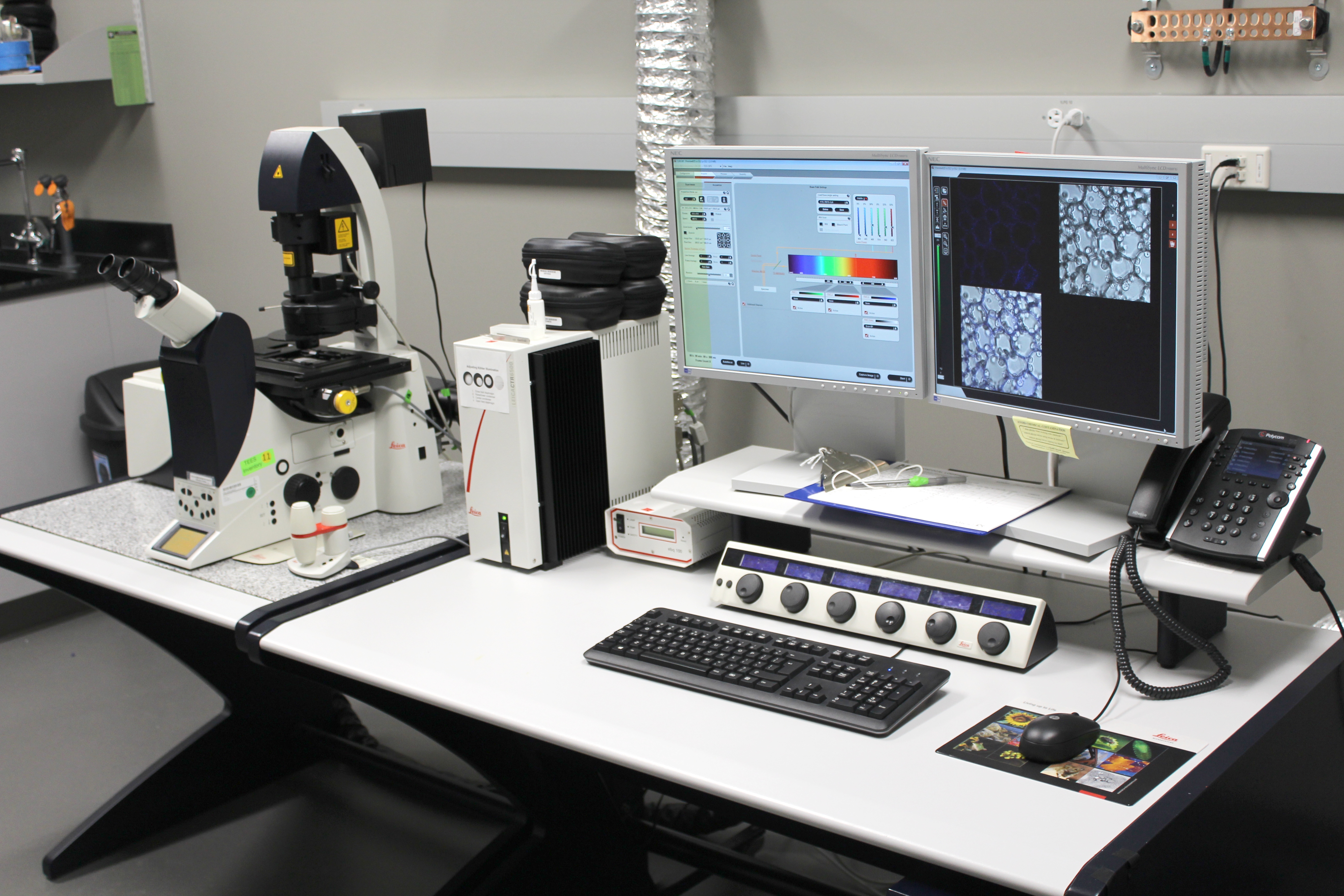
The Leica TCS SP5 microscope utilizes an inverted DMI 6000 microscope which is equipped with a motorized xy stage and epifluorescence illumination. Filter cubes for blue and green excitation (I3, N2.1) are available for visual inspection and focusing of samples. The conventional scanner has one transmitted light detector and three reflected/fluorescence detectors. The excitation laser lines available are 458, 476, 488, 514, 543 and 633 nm and the microscope is equipped with 10x, 40x and 63x dry objectives and a 63x oil immersion objective.

Confocal microscope instructions (updated 8/27/2010)

Key Features:

  • inverted DMI 6000 microscope with a motorized xy stage and epifluorescence illumination
  • 1 transmitted light, 3 reflected/fluorescence PMTs
  • excitation laser lines: 458, 476, 488, 514, 543 and 633 nm
  • 10x, 40x and 63x dry objectives, and 63x oil immersion objective
  • high resolution imaging 8k x 8k pixels
  • FRAP, FRET measurements
  • ROI scan
  • spectral un-mixing, deconvolution, colocalization
  • 3-D visualization

To be trained on this instrument, please contact Dr. Yordanos Bisrat.

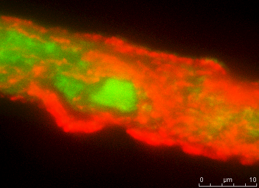
CdSe quantum dots (red) coating a protein fiber (green). 3D projection of an image stack taken with the Leica TCS SP5 laser confocal microscope using sequential scanning with excitation wavenlengths 488 nm (EGFP tagged protein fiber emitting at 507 nm) and 458 nm (CdSe emitting at 580 nm) using a 63x objective (dry, N.A. 0.7). Image taken by Ravish Majithia.
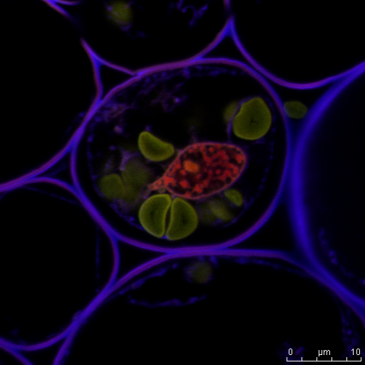
Convallaria 488 514 633 5x zoom

Primary Sidebar

Instruments

PI 85 and PI 95 PicoIndenter

AFM-IR

Omicron XPS/UPS

Nanoindenter

LYRA FIB-SEM

Ellipsometer

Spectrofluorometer

UV-Vis-NIR Spectrophotometer

Electopolishers

Confocal Microscope

FE-SEM

Cameca Ion Microprobe

Dimension Icon AFM

LYRA FIB-SEM

Fume Hoods

FERA FIB-SEM

Electron Microprobe

FTIR Spectrometer

Differential Scanning Calorimeter (DSC)

Thermal Mechanical Analysis (TMA)

Hot Disc Thermal Conductivity Analyzer

Dielectric Spectrometer

Tensile Stages (500 N and 10 kN)

Prep Lab

Titan Themis 300 S/TEM

Optical Microscope

PIPS II

SEM and TEM Plasma Cleaners

Particle Size Analyzer

Surface Area Analyzer

Footer

Materials Characterization Facility (MCF)

Frederick E. Giesecke Engineering Research Building
1617 Research Parkway
College Station, TX 77843

Texas A&M Experiment Station
  • Accessibility
  • State Links and Policies
  • Privacy Notice
  • Website Feedback
  • Texas A&M University

Copyright © 2025 · Texas A&M Engineering Experiment Station · All Rights Reserved