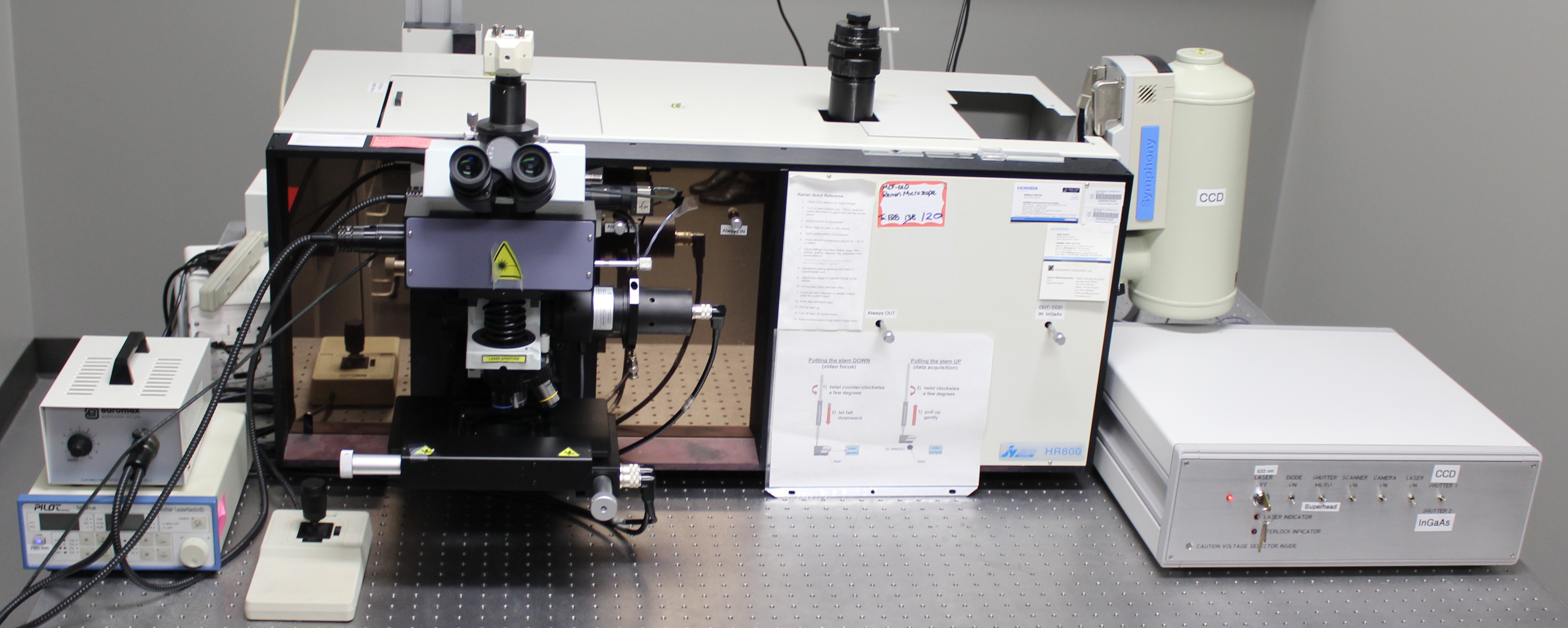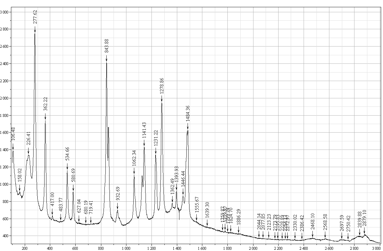
The Horiba Jobin-Yvon LabRam HR is a bench top confocal Raman system. The instrument provides highly specific spectral fingerprints which enables precise chemical and molecular characterization and identification. The raman microscope is based around the established high performance of the LabRam series of Raman microscopes, offering optimal confocal spatial and depth discrimination down to 1 μm, multiple laser options and automated XYZ mapping.
For more information please see: JY Horiba Raman Division
Microscope
Olympus BX 41 microscope. The computer-controlled motorized XYZ microscope stage with a step size 0.1 μm.
Raman module
The Raman module consists of a stigmatic 800 nm spectrograph with two confocal spectrometer entrances, one connected to the microscope, the other via a fiber optics coupler, and 633 nm and 785 nm laser excitation lines. The spectrometer is equipped with two detectors.
- Spectral Range: 400 nm – near-Infrared (NIR)
- Raman Shift: 100-6000 cm-1
- Spectral Resolution: depends on excitation frequency and grating used; Resolution = 0.16 cm-1 at 633 nm excitation with 1800 gr/mm and CCD detector; Resolution = 0.30 cm-1at 785 nm excitation with 1800 ln/mm grating and CCD detector
- Imaging Resolution: diffraction limited; 702 nm using 633 nm with 50x N.A. 0.55 objective; 870 nm using 785 nm with 50x N.A. 0.55 objective
- Detectors: (1) JY open electrode CCD with enhanced quantum efficiency in the spectral range 450 – 950 nm; (2) InGaAs diode array JY IGA-3000, with highest sensitivity between 900 nm and 1700 nm [InGaAs detector is currently not functioning]
**Please Note** Special Acknowledgement requirement for the new Raman/FTIR/AFM/NSOM: NSF guidelines mandate that the use of the funded Horiba Jobin Yvon/Nanonics Raman/FTIR/ AFM/NSOM must be properly acknowledged in any publications (including web pages): “The Raman/FTIR/AFM/NSOM acquisition was supported by the National Science Foundation under Grant No. BES-0421409”. Users are also required to file a copy of any relevant publication containing the acknowledgement with the MCF Administrative Office ([email protected]).
This instrument is currently not working. Please check the “Equipment Status” page for the change to “up”
To be trained on this instrument, please contact Dr. Jing Wu.

